
Your skin is easy to think of as a surface. You wash it, moisturize it, notice when it becomes dry or oily, and probably pay more attention when a pimple, rash, scar, or wrinkle appears.
But beneath that familiar surface is a remarkably complex organ.
Skin separates your internal environment from the outside world. It helps prevent water loss, protects against physical and chemical threats, participates in immune defense, regulates body temperature, allows you to sense touch and pain, supports hair and glands, and contributes to vitamin D production.
Understanding the layers of skin makes many everyday skincare questions easier to understand. Why does skin become dry? Why does a deeper cut bleed? Why does skin lose elasticity as we age? Why does the skin barrier matter? Why can repeated sun exposure affect both the appearance and health of your skin?
The answers often begin with anatomy.
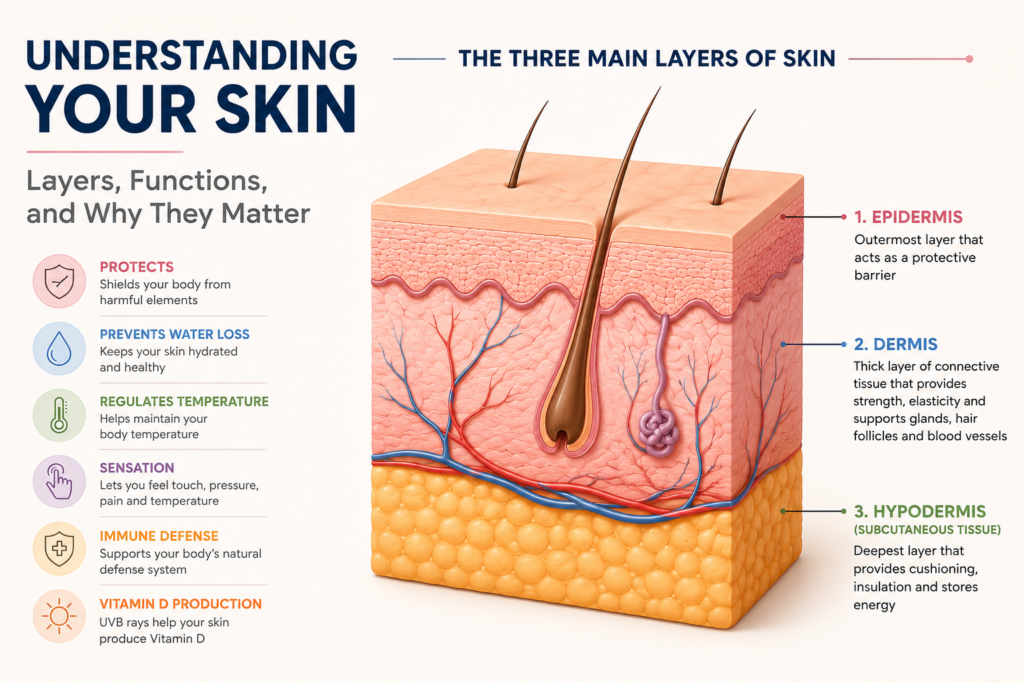
The skin is commonly described as having three major layers: the epidermis, dermis, and hypodermis, or subcutaneous tissue. Each has different structures and functions, but they work together as one interconnected system. The NCBI overview of skin anatomy describes the epidermis as the outer layer, the dermis as the deeper connective-tissue layer, and the subcutaneous tissue as the deeper layer containing fat and connective tissue.
Let’s look at what each layer does and why it matters for healthy skin.
What Are the Three Main Layers of Skin?
The three major layers commonly discussed in skin anatomy are:
| Skin layer | Location | Main functions |
|---|---|---|
| Epidermis | Outermost layer | Barrier protection, cell renewal, pigmentation and immune defense |
| Dermis | Beneath the epidermis | Strength, elasticity, sensation, blood supply, hair follicles and glands |
| Hypodermis | Beneath the dermis | Insulation, cushioning, energy storage and connection to deeper tissues |
There is a small anatomical distinction worth knowing. Some medical references describe the skin itself as consisting primarily of the epidermis and dermis, with the hypodermis located underneath rather than technically being part of the skin. Other educational resources commonly refer to the epidermis, dermis and hypodermis as the three main layers of skin. The NCBI Bookshelf’s anatomy reference uses the three-layer description, while also distinguishing the hypodermis as subcutaneous tissue.
For everyday skincare discussions, the three-layer model is useful because it shows how the surface of your skin connects with the deeper tissues underneath it.
1. The Epidermis: Your First Line of Defense
The epidermis is the outermost layer of skin. It is the part of your skin that directly interacts with the outside environment.
It may look simple from the outside, but the epidermis is a dynamic tissue that is constantly renewing itself. Its cells help create a protective barrier, contribute to skin pigmentation, participate in immune defense, and help control the movement of water between the body and the environment.
The primary cells of the epidermis are called keratinocytes. They are produced in the deeper part of the epidermis and progressively move toward the surface as they mature.
In most areas of the body, the epidermis contains four distinct layers. In areas of thick, hairless skin such as the palms and soles, it contains a fifth layer called the stratum lucidum. The five layers are:
- Stratum basale
- Stratum spinosum
- Stratum granulosum
- Stratum lucidum
- Stratum corneum
The NCBI’s detailed anatomy reference explains that the stratum lucidum is present in thick skin but absent from most areas of thinner skin.
Stratum Basale
The stratum basale, or basal layer, is the deepest part of the epidermis. This is where new keratinocytes are generated. The layer also contains melanocytes, which produce melanin, the pigment responsible for much of the color of human skin.
The basal layer therefore plays two important roles: maintaining epidermal renewal and contributing to pigmentation.
Stratum Spinosum
Above the basal layer is the stratum spinosum. It contains several layers of keratinocytes connected by structures called desmosomes. These connections help provide mechanical strength to the epidermis.
The layer also contains Langerhans cells, which participate in immune surveillance within the skin.
Stratum Granulosum
The stratum granulosum is where keratinocytes undergo further changes as they move toward the surface. Cells in this layer contribute to the development of the skin’s protective barrier. Lipids released into the spaces between cells become particularly important for limiting the movement of water through the epidermis.
Stratum Lucidum
The stratum lucidum is a thin, translucent layer found only in thick skin. You find this type of skin primarily on the palms of the hands and soles of the feet, where the epidermis experiences considerable friction. It sits between the stratum granulosum and stratum corneum.
Stratum Corneum
The stratum corneum is the outermost portion of the epidermis and is central to the skin’s barrier function. It consists largely of flattened, terminally differentiated cells called corneocytes, surrounded by an organized lipid matrix.
A useful way to visualize this structure is as a brick wall. The corneocytes are somewhat like bricks, while lipids between the cells act like mortar. The analogy isn’t perfect, but it captures an important concept: the structure is designed to create a relatively effective barrier between your body and the outside environment.
Research reviewed in the National Library of Medicine’s discussion of the skin’s defensive barrier identifies the stratum corneum as a critical component of the epidermal permeability barrier.
Why the Skin Barrier Matters
“Skin barrier” has become one of the most frequently used terms in modern skincare, and there is good biology behind it.
The barrier helps limit transepidermal water loss, or TEWL. In simple terms, it helps keep water inside the body while limiting the movement of unwanted substances from the environment into the skin. The stratum corneum isn’t simply a layer of dead cells. Its barrier properties depend on the organization of cells, lipids, proteins and other components.
Research describes the intercellular lipid matrix as containing important lipids such as ceramides, cholesterol and free fatty acids. These lipids help create a structure that restricts the outward movement of water. The National Library of Medicine’s review of skin barrier function explains this relationship in detail.
A more recent review in the National Library of Medicine’s PubMed Central database likewise describes the stratum corneum as the primary physical barrier and discusses its highly organized lipid structure.
This is why repeatedly irritating the skin can be counterproductive. The goal of skincare isn’t simply to remove as much from the surface as possible. Healthy skin needs its barrier to function properly.
2. The Dermis: Strength, Elasticity, Sensation and Support
Beneath the epidermis is the dermis.
If the epidermis is primarily associated with the skin’s external barrier, the dermis can be thought of as the structural and functional support layer underneath it.
The dermis contains:
- Collagen
- Elastin
- Blood vessels
- Lymphatic vessels
- Nerve structures
- Hair follicles
- Sweat glands
- Sebaceous glands
- Connective tissue
- Fibroblasts and other cells
According to the NCBI’s anatomy reference on the skin, the dermis has two major regions: the papillary dermis and the reticular dermis.
Papillary Dermis
The papillary dermis is the upper portion of the dermis, immediately beneath the epidermis. It is composed primarily of relatively loose connective tissue and forms the interface between the epidermis and deeper dermal structures. Small blood vessels in this region help support the surrounding tissues, while sensory structures contribute to the skin’s ability to detect changes in its environment.
Reticular Dermis
The reticular dermis is the deeper and generally thicker portion of the dermis. It contains denser connective tissue and substantial amounts of collagen and elastic fibers. This is particularly important when discussing skin strength, firmness and elasticity.
Collagen and Elastin: Why They Matter for Skin Appearance
Two words appear repeatedly in conversations about skin aging: collagen and elastin.
They aren’t interchangeable. Collagen provides structural strength. Elastin contributes to the skin’s ability to stretch and recoil. The dermal extracellular matrix contains a complex network of proteins and other molecules that determine many of the skin’s mechanical properties.
As skin ages, this structure changes. Collagen production, organization and degradation are affected, while elastic fibers can become altered as well. These changes contribute to changes in firmness, elasticity and wrinkle formation. Sun exposure can accelerate this process.
A 2026 review by researchers from Johns Hopkins University School of Medicine, published in Annals of Dermatology, describes chronic sun exposure as the most important external contributor to photoaging, which commonly presents with wrinkles and pigmentary changes. The 2026 review on photoaging is available through PubMed.
Earlier experimental research has also demonstrated that UV exposure can reduce the production of type I procollagen, an important structural component of skin. The findings are described in this PubMed-indexed research study on UV exposure and collagen.
The practical implication is straightforward: some skin aging is unavoidable, but cumulative environmental damage, particularly from UV exposure, is not something you have to ignore.
The Dermis Helps You Feel the World Around You
Your skin is also a sensory organ. Specialized nerve structures allow you to detect sensations such as:
- Touch
- Pressure
- Temperature
- Pain
This sensory function is protective. If you touch a very hot surface, pain and temperature receptors help alert you to the danger. If something presses against your skin, sensory information tells your nervous system what is happening.
The NCBI’s overview of skin anatomy describes the skin as a sense organ containing specialized sensory structures involved in detecting touch, temperature and pain.
3. The Hypodermis: Cushioning, Insulation and Energy Storage
Below the dermis lies the hypodermis, also called the subcutaneous tissue. The hypodermis contains a significant amount of adipose tissue, along with connective tissue, blood vessels and nerves.
Its main functions include:
- Insulating the body
- Storing energy
- Absorbing and distributing mechanical forces
- Connecting the skin to underlying tissues
- Allowing movement between superficial and deeper structures
The amount of subcutaneous fat isn’t the same everywhere. It varies by body region, age, genetics, sex and other factors.
The NCBI Bookshelf description of skin anatomy identifies the hypodermis as the deeper layer containing adipose tissue and connective tissue beneath the dermis.
This layer is also why a person’s skin isn’t simply attached directly to the underlying muscle or bone. It provides a flexible transition between the superficial skin and deeper structures.
What Are the Main Functions of Skin?
Knowing the layers is useful, but understanding what the skin actually does puts the anatomy into perspective.
1. Protection
The skin provides a physical and biological barrier between your body and the environment. It helps defend against mechanical injury, chemicals, microorganisms and UV radiation.
The epidermis provides much of the physical barrier, while deeper tissues and immune mechanisms contribute additional protection. The NCBI’s review of skin anatomy and function describes protection against trauma, pathogens, toxins and ultraviolet radiation as among the skin’s major roles.
2. Prevention of Excessive Water Loss
Your body continuously loses some water through the skin. The epidermal permeability barrier helps keep this loss under control.
The stratum corneum is particularly important because its structure limits the movement of water through the outermost layer. When this barrier is disrupted, water loss can increase.
This is one reason compromised skin can feel dry, rough or irritated.
3. Temperature Regulation
Skin participates in thermoregulation.
Blood vessels in the dermis can change their diameter, influencing how much heat is transferred between the body and environment. Sweat glands also produce sweat, which can cool the body as it evaporates.
The NCBI’s skin anatomy reference describes the skin’s blood supply as an important component of temperature regulation.
4. Sensation
The skin constantly gathers information about the external environment. Touch, pressure, pain and temperature are detected through sensory structures associated with the skin and nervous system.
This isn’t a secondary function. Sensation helps you avoid injury and interact with your surroundings safely.
5. Immune Defense
The skin is an active part of the body’s immune system. Physical barriers limit the entry of microorganisms, while specialized cells and other immune mechanisms respond to threats that reach the tissue.
The relationship between barrier function and immunity is particularly important in inflammatory skin disorders. Research on the defensive barrier function of skin describes the skin as an important primary defense system with antimicrobial and immune-related functions.
6. Vitamin D Production
The skin also participates in vitamin D synthesis.
When UVB radiation reaches uncovered skin, it can convert a precursor called 7-dehydrocholesterol into previtamin D3, which subsequently undergoes further conversion to vitamin D3.
However, this doesn’t mean that intentionally seeking prolonged unprotected sun exposure is a good strategy for maintaining vitamin D.
The amount of vitamin D produced from sunlight varies depending on factors such as season, time of day, geographic conditions, cloud cover, skin pigmentation and sunscreen use. The NIH Office of Dietary Supplements’ vitamin D fact sheet explains these variables and the uncertainty around how much sun exposure is needed for adequate vitamin D production.
How the Skin Layers Work Together
The most useful way to understand skin anatomy is not to think of the epidermis, dermis and hypodermis as three independent layers.
They are interconnected. Consider what happens when you cut your finger. A superficial injury may damage mainly the epidermis. A deeper injury can extend into the dermis, where blood vessels and nerves are located. If an injury extends further into subcutaneous tissue, it can affect fat and deeper structures.
This helps explain why the depth of an injury matters. The epidermis itself does not contain blood vessels. It receives nutrients and oxygen from blood vessels in the underlying dermis. Once an injury reaches the dermis, it can damage those vessels and cause bleeding.
The body then initiates a coordinated wound-healing response involving processes such as clot formation, inflammation, tissue formation and remodeling. So even a small wound can demonstrate how closely the different layers work together.
How Skin Changes as You Age
Skin changes throughout life.
The rate of cell renewal can change. The dermal extracellular matrix changes. Collagen and elastic fibers are altered. Blood vessels and glands can change. Wound healing can become less efficient, and the distribution of subcutaneous fat can change as well.
The NCBI’s review of skin anatomy notes that skin thickness varies by body site and changes with age and other biological factors.
Skin aging is often divided conceptually into two broad categories. Intrinsic aging refers to the natural biological aging process. Extrinsic aging refers to changes influenced by environmental exposures and lifestyle factors.
UV exposure is particularly important because chronic exposure can contribute to photoaging. The recent 2026 review on photoaging describes chronic sun exposure as the major external contributor to this process.
This distinction matters because you cannot stop intrinsic aging. You can, however, reduce some forms of avoidable environmental damage.
Why Sun Protection Matters Beyond Sunburn
Sunscreen is sometimes viewed as something you need only when you’re going to the beach or spending several hours outdoors.
The biology is more complicated. UV radiation can damage cells and contribute to changes in the skin’s connective tissue. Over time, this cumulative exposure contributes to photoaging and increases the risk of skin cancer.
The American Academy of Dermatology recommends selecting a sunscreen that offers broad-spectrum protection, SPF 30 or higher, and water resistance. Its official sunscreen guidance explains these recommendations.
Sun protection also shouldn’t be reduced to sunscreen alone.
Shade, protective clothing and avoiding excessive UV exposure can all contribute to reducing cumulative exposure. The AAD’s sun-protection guidance discusses these measures alongside sunscreen.
What Skin Anatomy Means for Your Skincare Routine
Understanding the layers of skin doesn’t mean you need a complicated skincare routine. In fact, it can have the opposite effect.
Once you understand what the skin is trying to do naturally, many skincare principles become easier to understand.
Protect the Skin Barrier
The outer barrier is doing important work every minute. Repeatedly exposing skin to irritating products, excessive cleansing or other forms of unnecessary stress can interfere with normal barrier function.
This doesn’t mean you should never exfoliate or use active ingredients. It means that irritation isn’t automatically a sign that a product is “working.” If a routine repeatedly leaves your skin uncomfortable, excessively dry or irritated, the answer isn’t necessarily to add another product.
Moisturize When Your Skin Needs It
Moisturizers can help reduce dryness and improve the skin’s hydration. For people with dry skin, applying moisturizer after cleansing can help maintain hydration. The best formulation depends on factors such as skin type, climate and personal tolerance.
There isn’t one universally perfect moisturizer because healthy skincare isn’t identical for everyone.
Protect Your Skin From Excessive UV Exposure
UV protection is one of the clearest connections between skin anatomy and everyday skincare. UV exposure affects both the epidermis and deeper structures involved in skin aging.
Using a broad-spectrum sunscreen with SPF 30 or higher is one practical step recommended by the American Academy of Dermatology.
Don’t Assume More Products Mean Better Skin
A longer routine isn’t automatically a better routine. If five products work well together and you can use them consistently, that’s useful. If ten products repeatedly irritate your skin, the additional complexity may be doing more harm than good.
Your skin is already a sophisticated biological system. Skincare should generally support that system rather than constantly trying to overpower it.
Common Misconceptions About the Layers of Skin
“The epidermis is just dead skin.”
That’s an oversimplification.
The outermost stratum corneum contains predominantly dead, terminally differentiated cells, but the epidermis as a whole contains living cells and continuously renews itself.
The deeper epidermal layers contain active cells responsible for generating and maintaining the tissue.
“The hypodermis is just fat.”
Fat is a major component of the hypodermis, but it isn’t the only component.
The tissue also contains connective tissue, blood vessels, nerves and other structures. It contributes to insulation, cushioning and attachment of the skin to deeper tissues.
“The skin barrier is only the stratum corneum.”
The stratum corneum is central to the physical permeability barrier, but barrier function involves more than one mechanism.
The skin also has chemical, immune and biological defenses. Modern research increasingly treats the barrier as an interconnected system rather than a simple wall.
“Wrinkles are caused only by dry skin.”
Dryness can make fine lines appear more noticeable, but wrinkles have a deeper biological basis.
Changes in collagen, elastic fibers and other components of the dermis contribute to structural skin aging. UV exposure can accelerate these changes through photoaging.
“Darker skin cannot be damaged by the sun.”
Melanin provides some natural protection against UV-related damage, but it does not make skin immune to sunburn, photoaging or skin cancer.
The NCBI’s skin assessment reference notes that darker skin contains more melanin and generally has greater protection from UV-induced damage, while also making clear that darker skin can still experience sunburn.
Frequently Asked Questions
What are the three layers of skin?
The three layers commonly described are the epidermis, dermis and hypodermis. The epidermis forms the outer protective layer, the dermis contains connective tissue, blood vessels, nerves, hair follicles and glands, and the hypodermis contains substantial amounts of fat and connective tissue beneath the dermis.
Which layer of skin is responsible for the skin barrier?
The stratum corneum, the outermost portion of the epidermis, is a major component of the skin’s permeability barrier. Its corneocytes and organized lipid matrix help limit water loss and protect the body from external threats.
Which layer contains collagen?
The dermis contains much of the skin’s collagen and elastic connective tissue. These components provide structural support, strength and elasticity.
Why does skin lose elasticity with age?
Skin aging involves changes in the dermal extracellular matrix, including collagen and elastic fibers. Intrinsic aging contributes to these changes, while environmental factors such as chronic UV exposure can accelerate them.
Does the skin produce vitamin D?
Yes. UVB radiation can initiate vitamin D synthesis in the skin. However, production varies considerably depending on factors including season, time of day, skin pigmentation and geographic conditions.
Why does a deep cut bleed while a superficial scratch may not?
Blood vessels are located in the dermis rather than the epidermis. An injury that reaches the dermis can therefore damage blood vessels and cause bleeding, while a very superficial injury may affect only the epidermis.
Is the hypodermis technically part of the skin?
This depends on the anatomical terminology being used. Many references describe the skin as consisting of the epidermis and dermis and place the hypodermis beneath them as subcutaneous tissue. Other educational references use a three-layer model that includes the hypodermis. The distinction is mainly about anatomical classification rather than the functions of the tissue itself.
Can skincare products reach the deeper layers of skin?
Most topical products primarily interact with the outer layers of the skin, and the stratum corneum represents a significant barrier to penetration. How deeply a substance can penetrate depends on its chemical properties, formulation, concentration, skin condition and the route of delivery. Research on skin barriers and dermal drug delivery explains why the skin’s structure creates significant barriers to penetration.
The Bottom Line
Your skin is much more than the surface you see in the mirror.
The epidermis forms the body’s first major external barrier and continuously renews itself. The dermis provides structural support and contains collagen, elastic fibers, blood vessels, nerves, hair follicles and glands. The hypodermis provides cushioning, insulation, energy storage and a connection between the skin and deeper tissues.
Together, these layers allow skin to perform an impressive range of functions: protecting the body, limiting water loss, regulating temperature, detecting sensations, participating in immune defense and contributing to vitamin D production.
Understanding this structure also puts skincare into perspective.
Healthy skin doesn’t necessarily require an elaborate routine. The fundamentals are much simpler: support the skin barrier, avoid unnecessary irritation, moisturize when appropriate, and reduce excessive UV exposure.
The deeper lesson is that your skin is already an active biological system. Good skincare isn’t about treating it like an inert surface. It’s about understanding how that system works and making choices that support its ability to protect, repair and maintain itself.